Understanding Laser Photocoagulation for Age-Related Macular Degeneration (AMD)
Laser photocoagulation is a type of laser surgery for the eyes. It's done to treat age-related macular degeneration (AMD). AMD is a condition that can lead to loss of eyesight, especially in your central vision. Laser photocoagulation uses a laser to seal off abnormal blood vessels in your eye. Laser photocoagulation can’t restore eyesight that you have already lost. However, it may slow down the damage to your central vision.
What is AMD?
The retina is a very thin layer of cells in the back of your eye. It converts light into electrical signals and then sends these signals to your brain. The macula is the sensitive center part of your retina. This area gives you detailed vision in the middle of your visual field. AMD damages your macula. The macula may become thinner, and deposits of material can develop behind it. Blood vessels may start growing beneath your macula. This can cause fluid to leak beneath your macula. This excess fluid can lead to eyesight loss.
AMD has 2 types: dry and wet. Abnormal blood vessel growth is present in only the wet type. AMD is a common cause of severe eyesight loss in older adults. In rare cases, it can result in total blindness. AMD affects the macula so you may still have your side (peripheral) vision. Loss of central vision can be gradual or sudden.
Why laser photocoagulation for AMD is done
Laser photocoagulation is 1 type of treatment for AMD. It's a choice only for certain people with wet AMD. Your healthcare provider may advise the surgery if your abnormal blood vessels are close together. The surgery may be less effective if you have scattered blood vessels or vessels located in the center of the macula. Your healthcare provider may be more likely to advise the surgery if your eyesight loss happens suddenly instead of slowly.
AMD can also be treated with medicine that is injected into the eye and slows down the growth of abnormal blood vessels. Your healthcare provider may use both laser photocoagulation and injected medicine to treat your AMD.
How laser photocoagulation for AMD is done
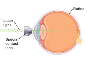
The eye care provider will use eye drops to enlarge (dilate) your pupil. Your pupil will stay dilated for several hours after the surgery. The eye care provider will put a special type of contact lens into the affected eye. This lens helps focus a beam of laser light on the retina using a tool called a slit lamp. The eye care provider uses the laser to seal off the abnormal blood vessels beneath the macula.

Risks of laser photocoagulation
During laser photocoagulation, the eye care provider burns away part of the macula. You might have a blind spot where the laser creates a scar. In some cases, this eyesight loss might be worse than the eyesight loss from not treating the eye. This is something to talk with your eye care provider about when considering the surgery.
The surgery has some other risks, such as:
-
Accidental treatment of the central macula, which causes a worse blind spot
-
Bleeding into the eye
-
Damage to the retina from the laser scar, right away or years later
-
Abnormal blood vessels growing back
-
Need for repeat treatment
Your risks may differ according to your age, your general health, and the type of your AMD. Ask your healthcare provider which risks apply most to you.